
You’re 52. You’re Losing Bone Right Now. No One is Measuring It.
During the premenopausal and early perimenopausal years, bone mineral density barely changes. The skeleton is stable.

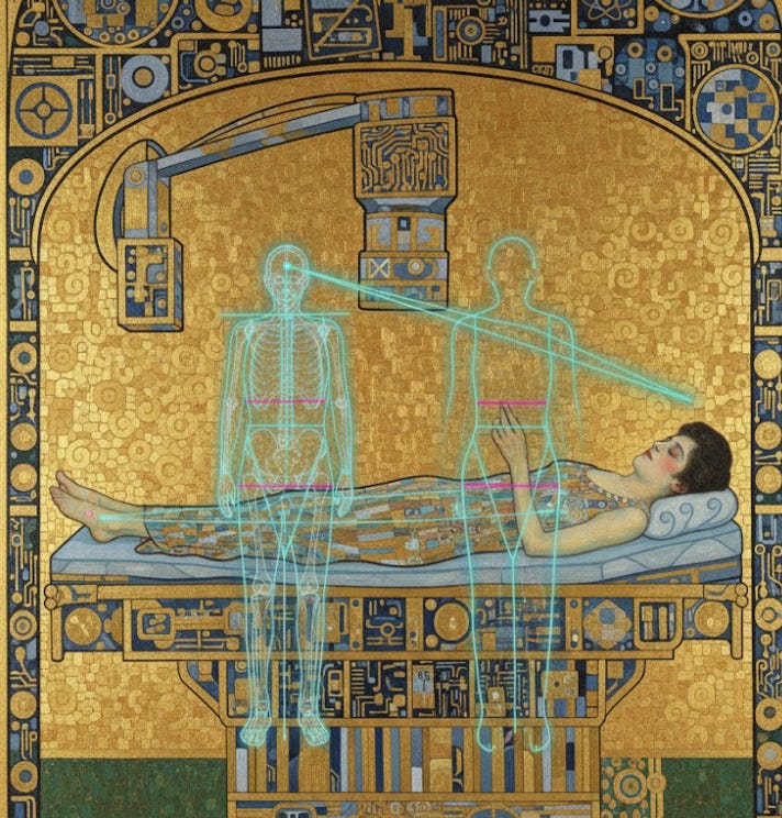
Know Your Numbers, Trust Your Body
She’s 52. Active. Runs three miles twice a week. Eats well. Takes a multivitamin. She mentioned to her gynecologist that her mother broke a hip at 72. She asked if she should get a bone density scan.
Her doctor said no. Not yet. Guidelines say 65.
So she went home. Reassured. She’ll think about it again in 17 years.
By then, she may have lost up to 20% of her bone mineral density. The first fracture will be her screening test.
This is how we manage bone health in women. We wait until the damage is done, call it prevention, and act surprised when bones break.
The Biology Your Doctor Should Know
The Study of Women’s Health Across the Nation, known as SWAN, is the largest and most rigorous longitudinal investigation of the menopausal transition ever conducted. Over 3,000 women across five ethnic groups, followed for more than 20 years at five clinical centers. It remains the definitive dataset on what actually happens to women’s bodies during this transition [1].
Here is what SWAN found about bone:
During the premenopausal and early perimenopausal years, bone mineral density barely changes. The skeleton is stable. Then, in the late perimenopause, bone loss accelerates dramatically. In the lumbar spine, women lose 1.8 to 2.3% per year. In the hip, 1.0 to 1.4% per year [1].
These are not small numbers.
If that rate continues for five years, and it often does, the average woman’s spine loses 7 to 10% of its mineral density. The hip loses 5 to 7%. Those losses translate directly into fracture risk: a 50 to 100% increase [1].
More recent SWAN analyses identified what researchers call the “transmenopause,” a critical 3-year window spanning from one year before the final menstrual period to two years after it. During this window, White women lose an average of 2.5% per year in the lumbar spine and 1.8% per year in the femoral neck. Before this window, there is no appreciable bone loss at either site [2].
The Endocrine Society estimates that up to 20% of a woman’s total bone mass can be lost during the menopausal transition [3]. Women lose approximately 50% of their trabecular bone and 30% of their cortical bone over a lifetime, and roughly half of that loss occurs in the first 10 years after menopause [1].
The average age of perimenopause onset is 47.5 years [4]. The average age of menopause in the United States is 51. The rapid bone loss phase begins around age 50 for most women.
The USPSTF recommends screening with DXA at age 65.
That is a 15-to-18-year gap between when bone loss begins and when anyone looks.
What the Guidelines Actually Say
In January 2025, the US Preventive Services Task Force updated its osteoporosis screening recommendation. The core advice has not changed since 2011 [5]:
Screen all women 65 and older with DXA. That’s a B recommendation, meaning moderate certainty of moderate net benefit.
For postmenopausal women younger than 65, the USPSTF recommends screening only if they are “at increased risk,” determined by a clinical risk assessment tool such as FRAX. That’s also a B recommendation [5].
For perimenopausal women, there is no recommendation at all. Not a recommendation against screening. Not insufficient evidence. Simply silence.
The problem is not that these guidelines are wrong. The problem is what they miss. A 48-year-old woman in late perimenopause who is losing 2.5% of her spinal bone density per year does not fit the screening algorithm. She is not postmenopausal. She is not 65. She has no fracture history. FRAX, which begins at age 40, was designed primarily to predict fractures in older postmenopausal populations. Its accuracy in younger women is moderate at best [5].
So the fastest period of bone loss in a woman’s life falls neatly into a guideline gap. The biology is screaming. The guidelines are silent.
The Numbers That Should Terrify Us
One in two White women will experience an osteoporotic fracture in their lifetime. For White women, the lifetime risk of hip fracture alone is 1 in 6. That is more common than a breast cancer diagnosis, which is 1 in 9 [6].
We screen aggressively for breast cancer starting at 40. We screen for osteoporosis starting at 65. The disease with the higher lifetime risk gets 25 fewer years of surveillance.
Hip fractures are not minor injuries. Mortality rates in the first year after a hip fracture range from 20 to 24% [7]. Only 30 to 40% of patients recover their previous level of function. One third of formerly independent, community-dwelling women end up in nursing homes within a year of the fracture [7]. The annual economic burden of osteoporotic fractures in the United States exceeds $17.9 billion [6].
And here is the number that should change practice: a 10% loss of bone mass in the hip results in a 2.5 times greater risk of hip fracture [8]. That 10% is not a hypothetical. It is what many women lose during the menopausal transition.
The 2025 USPSTF report itself acknowledges that 27.1% of women 65 and older have osteoporosis [5]. Which means more than one in four women already have the disease by the time we first look.
That is not early detection. That is late discovery.
Know Your Numbers: What to Ask For and When
This is the “Know Your Numbers” part. The part your doctor may not volunteer.
1. Know Your FRAX Score
The Fracture Risk Assessment Tool is free and takes two minutes. You can calculate it yourself at frax.shef.ac.uk. It estimates your 10-year probability of a major osteoporotic fracture and a hip fracture using clinical risk factors alone, without a DXA scan [9].
The inputs: your age, sex, weight, height, prior fracture, parental hip fracture, smoking status, alcohol use, glucocorticoid use, rheumatoid arthritis, and secondary osteoporosis causes.
A 10-year major osteoporotic fracture risk of 9.3% or higher, which is the risk level of an average 65-year-old White woman, is the USPSTF benchmark at which DXA screening becomes recommended [5]. Many women in their late 40s and early 50s with even one or two risk factors will meet that threshold. They just have to know to check.
2. Request a Baseline DXA During Perimenopause
The official guideline says 65. But the USPSTF also says that postmenopausal women younger than 65 with risk factors should be screened. And the Menopause Society states that women with established risk factors for osteoporosis should consider bone density assessment starting at menopause [10].
Risk factors that warrant earlier screening: a parent who broke a hip, low body weight (under 127 pounds), current smoking, excessive alcohol, early menopause (before 45), prolonged amenorrhea before menopause, glucocorticoid use, rheumatoid arthritis, type 1 diabetes, or hyperthyroidism.
If you have any of these, request a DXA scan. If your provider says it’s not indicated, ask them to document their refusal. A baseline DXA at the onset of menopause gives you something invaluable: a starting point. Without it, you will never know how much bone you have lost until you have lost enough to fracture.
3. Know Your 25-Hydroxyvitamin D Level
The SWAN Bone Study found that women with serum 25(OH)D levels below 20 ng/mL had significantly increased fracture risk during the menopausal transition. Higher levels were associated with less bone loss [11].
The Institute of Medicine defines adequacy as 20 ng/mL or above. Many bone health experts recommend at least 30 ng/mL for optimal skeletal protection [12].
This is a simple blood test. You should know your number. If it is below 20, you are deficient and need aggressive supplementation. If it is between 20 and 30, there is evidence that you would benefit from supplementation. The daily recommendation for most adults is 600 to 800 IU of vitamin D3, but many bone health experts recommend 1,000 to 2,000 IU daily, especially during and after the menopausal transition.
4. Know Your Calcium Intake
Postmenopausal women need 1,200 mg of calcium daily. Premenopausal women need 1,000 mg. Most American women get about 600 mg from their diet [13].
Do not guess. Track it for three days. One cup of milk or yogurt provides about 300 mg. One cup of cooked kale about 180 mg. Fortified orange juice about 350 mg per cup.
If you are falling short, dietary sources are preferable to supplements. If supplements are needed, take no more than 500 to 600 mg at a time for better absorption, and take them with food.
5. Know Your Exercise Prescription
This is not “stay active.” This is specific.
SWAN data showed that women who increased their leisure-time physical activity from early perimenopause to late perimenopause had significantly slower rates of BMD loss [14]. The data support weight-bearing and resistance exercise, not just walking.
The specific prescription: weight-bearing impact exercise (walking, jogging, dancing, stair climbing) for at least 30 minutes most days, plus resistance training (targeting major muscle groups) at least two to three times per week. Balance training becomes increasingly important to prevent falls.
The Role of Hormone Therapy
This deserves direct language. Menopausal hormone therapy is the most effective intervention for preventing bone loss during the menopausal transition.
The Women’s Health Initiative demonstrated a 34% reduction in fracture risk with hormone therapy, even in women at low baseline risk [15]. The International Menopause Society’s 2024 White Paper describes MHT as “foundational” for bone health maintenance, particularly when initiated in early postmenopause and in women under 60 [16]. It reduces bone turnover, increases bone mineral density, and lowers fracture risk at all skeletal sites.
The FDA has approved estrogen therapy specifically for the prevention of osteoporosis in postmenopausal women.
Yet many women who would benefit from hormone therapy during the critical bone loss window are never offered it. And many physicians, still operating under outdated fears from the early WHI interpretation, actively discourage it. The result: women lose bone during the exact years when the most effective prevention exists, and are told to wait for a DXA scan 15 years later.
Hormone therapy is not appropriate for every woman. But the conversation about its bone-protective effects should happen with every woman entering the menopausal transition. Waiting until 65 to discuss bone health means the opportunity for prevention has already passed.
This Is Not Conservative Medicine
Let me be clear about what we are doing.
We know that bone loss accelerates dramatically in the late perimenopause. The SWAN study has documented this across more than 2,000 women over more than two decades. The biology is not in dispute.
We know that the rapid bone loss phase begins, on average, around age 50 and spans a critical 3-year window around the final menstrual period. We know that by the time most women are screened at 65, up to 20% of their bone mass may already be gone.
We know that hip fractures kill 20 to 24% of women in the first year. We know that the lifetime risk of hip fracture in White women exceeds the lifetime risk of breast cancer. We know that a 10% loss of bone in the hip more than doubles fracture risk.
We know that FRAX is free and takes two minutes. We know that vitamin D levels are a simple blood test. We know that DXA is a low-cost, low-radiation scan that takes 15 minutes. We know that hormone therapy reduces fracture risk by a third, that bisphosphonates reduce vertebral fractures by half, and that exercise during perimenopause slows bone loss.
We know all of this.
And we wait until 65.
We do not wait until 65 to screen for breast cancer. We do not wait until 65 to check cholesterol. We do not wait until 65 to monitor blood pressure or blood sugar. For every other major chronic disease, we screen during the years when intervention can change the trajectory. For osteoporosis, we screen after the trajectory is set.
This is not careful medicine. This is not conservative medicine. This is the medical system choosing, year after year, to let women lose bone in the dark.
It does not have to be this way. You can calculate your FRAX score today. You can ask for a vitamin D level at your next visit. You can request a baseline DXA when you enter the menopausal transition. You can start the conversation about hormone therapy before the window closes.
Your bones are not waiting until 65. Your doctors should not be either.
If this post changed how you think about bone health, share it with a friend who is in her 40s or 50s. This is the information that should be in every gynecologist’s office, and it is not.
Subscribe to ObGyn Intelligence for evidence-based women’s health that challenges the status quo.
References
Finkelstein JS, Brockwell SE, Mehta V, et al. Bone mineral density changes during the menopause transition in a multiethnic cohort of women. J Clin Endocrinol Metab. 2008;93(3):861-868.
Karlamangla AS, Burnett-Bowie SM, Engelman CD, et al. Bone health during the menopause transition and beyond. Obstet Gynecol Clin North Am. 2018;45(4):695-708.
Endocrine Society. Menopause and Bone Loss. Endocrine Society Patient Education. 2022.
Gold EB. The timing of the age at which natural menopause occurs. Obstet Gynecol Clin North Am. 2011;38(3):425-440.
US Preventive Services Task Force. Screening for osteoporosis to prevent fractures: US Preventive Services Task Force recommendation statement. JAMA. 2025;333(6):498-509.
International Osteoporosis Foundation. Epidemiology of osteoporosis and fragility fractures. IOF Facts and Statistics. Updated 2024.
Vidal EI, Moreira-Filho DC, Pinheiro RS, et al. Mortality after osteoporotic hip fracture: incidence, trends, and associated factors. J Orthop Surg Res. 2019;14(1):202.
Klotzbuecher CM, Ross PD, Landsman PB, et al. Patients with prior fractures have an increased risk of future fractures: a summary of the literature and statistical synthesis. J Bone Miner Res. 2000;15(4):721-739.
Schini M, Johansson H, Harvey NC, et al. An overview of the use of the fracture risk assessment tool (FRAX) in osteoporosis. J Endocrinol Invest. 2024;47(3):501-511.
The North American Menopause Society. Management of osteoporosis in postmenopausal women: the 2021 position statement of The North American Menopause Society. Menopause. 2021;28(9):973-997.
Cauley JA, Greendale GA, Ruppert K, et al. Serum 25 hydroxyvitamin D, bone mineral density and fracture risk across the menopause. J Clin Endocrinol Metab. 2015;100(5):2046-2054.
Bischoff-Ferrari HA, Giovannucci E, Willett WC, et al. Estimation of optimal serum concentrations of 25-hydroxyvitamin D for multiple health outcomes. Am J Clin Nutr. 2006;84(1):18-28.
National Institutes of Health Office of Dietary Supplements. Calcium: Fact Sheet for Health Professionals. Updated 2024.
Greendale GA, Huang MH, Cauley JA, et al. Leisure time physical activity and bone mineral density preservation during the menopause transition and postmenopause: the SWAN study. Lancet Reg Health Am. 2023;20:100481.
Cauley JA, Robbins J, Chen Z, et al. Effects of estrogen plus progestin on risk of fracture and bone mineral density: the Women’s Health Initiative randomized trial. JAMA. 2003;290(13):1729-1738.
Panay N, Anderson RA, Engel J, et al. Menopause and MHT in 2024: addressing the key controversies. An International Menopause Society White Paper. Climacteric. 2024;27(5):441-457.
Devastating breakdown of the guideline gap. The fact that we screen for breast cancer at 40 but osteoporosis at 65 when fractures have a higher lifetime risk is honestly wild. That 15-year window where women lose up to 20% bone density whiel nobody is measuring anything is textbook late discovery masquerading as prevention. The SWAN data is so cleartoo - why are we ignoring our own research?